
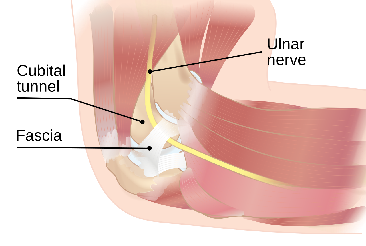
Overview — Cubital Tunnel Syndrome
Cubital tunnel syndrome (CuTS) is a compressive neuropathy of the ulnar nerve at the elbow. It is the second most common compression neuropathy of the upper limb after carpal tunnel syndrome. The ulnar nerve is vulnerable at the elbow because it passes through a narrow fibro-osseous tunnel behind the medial epicondyle and becomes stretched during elbow flexion.
Patients usually present with numbness and tingling in the little finger and ulnar half of the ring finger, often aggravated by prolonged elbow flexion, sleeping with the elbow flexed, leaning on the elbow, or using a phone. Advanced disease produces intrinsic hand weakness, reduced grip and pinch strength, Froment sign, Wartenberg sign, and clawing of the ring and little fingers.
- High-yield definition: Cubital tunnel syndrome = ulnar nerve compression at the elbow, most commonly beneath Osborne ligament, producing sensory symptoms in the ulnar one-and-a-half digits and intrinsic muscle weakness in severe disease.
- Early disease is predominantly sensory; late disease causes motor weakness and intrinsic wasting.
- Diagnosis is mainly clinical and may be confirmed with nerve conduction studies and electromyography.
Epidemiology
Cubital tunnel syndrome is less common than carpal tunnel syndrome but is the most common site of ulnar nerve entrapment. It is encountered frequently in orthopaedic, hand surgery, neurology, and occupational health practice. The condition may be idiopathic, post-traumatic, occupational, or associated with deformity around the elbow.
- Second most common upper-limb compression neuropathy after carpal tunnel syndrome.
- Most common site of ulnar nerve entrapment.
- Commonly affects adults involved in repetitive elbow flexion, prolonged computer or phone use, driving, cycling, or work requiring pressure on the elbow.
- Risk is increased in previous elbow fracture, cubitus valgus, elbow arthritis, ganglion, synovitis, diabetes mellitus, and occupations using vibrating tools.
- Bilateral disease can occur, but symptoms may be more prominent in the dominant or more exposed limb.
Ulnar Nerve Anatomy Relevant to Cubital Tunnel Syndrome
The ulnar nerve arises from the medial cord of the brachial plexus, carrying fibres mainly from C8 and T1. It descends along the medial arm, pierces the medial intermuscular septum, passes behind the medial epicondyle, and enters the forearm between the two heads of flexor carpi ulnaris. At the elbow, the nerve is superficial and lies close to bone, making it vulnerable to compression, traction, and direct trauma.
| Level | Anatomical Course | Clinical Importance |
|---|---|---|
| Arm | Runs medial to brachial artery, then pierces medial intermuscular septum | Arcade of Struthers may be a proximal compression site |
| Elbow | Passes posterior to medial epicondyle through cubital tunnel | Most common compression site; nerve stretches during elbow flexion |
| Forearm | Enters between two heads of flexor carpi ulnaris | FCU aponeurosis can compress the nerve distal to the tunnel |
| Wrist/Hand | Passes through Guyon canal superficial to flexor retinaculum | Differentiate cubital tunnel syndrome from ulnar tunnel syndrome at wrist |
- Exam pearl: ulnar nerve compression at the elbow may affect both FCU/FDP and intrinsic hand muscles, while compression at Guyon canal spares forearm muscles.
Cubital Tunnel Anatomy & Boundaries
The cubital tunnel is a fibro-osseous canal located posterior to the medial epicondyle. The ulnar nerve passes through this tunnel before entering the forearm. During elbow flexion, the tunnel volume decreases and the ulnar nerve is stretched, which explains why symptoms worsen when the elbow is kept flexed.
| Boundary / Structure | Anatomy | Clinical Significance |
|---|---|---|
| Roof | Osborne ligament / cubital tunnel retinaculum | Common compression structure; divided during decompression |
| Floor | Posterior bundle of medial collateral ligament and elbow capsule | Arthritis or osteophytes may narrow tunnel |
| Medial boundary | Medial epicondyle | Prominent epicondyle may contribute to traction/compression |
| Lateral boundary | Olecranon | Nerve lies between olecranon and medial epicondyle |
| Distal continuation | Deep flexor pronator aponeurosis / FCU aponeurosis | Must be released adequately during decompression |
Compression Sites Around the Elbow
Although the term cubital tunnel syndrome usually refers to compression beneath Osborne ligament at the elbow, the ulnar nerve may be compressed at multiple points around the elbow. A complete decompression requires awareness of all potential constriction sites.
| Site | Structure | Clinical/Surgical Importance |
|---|---|---|
| Arcade of Struthers | Fascial band in distal medial arm | Can cause proximal compression; release if tight during transposition |
| Medial intermuscular septum | Septum between anterior and posterior compartments | Can kink nerve after anterior transposition if not excised/released |
| Cubital tunnel | Osborne ligament | Most classical site of compression |
| FCU aponeurosis | Fascial arch between two heads of FCU | Important distal compression site; must be released |
| Deep flexor pronator fascia | Deep fascia distal to FCU | May cause persistent symptoms if missed |
Pathophysiology
The ulnar nerve may be injured by compression, traction, friction, or subluxation. During elbow flexion, the cubital tunnel cross-sectional area decreases, intraneural pressure increases, and the ulnar nerve elongates. Repeated or prolonged elbow flexion can therefore produce ischemia and conduction block. Chronic compression causes demyelination first, followed by axonal loss and denervation of intrinsic hand muscles.
- Core mechanism: elbow flexion narrows the cubital tunnel and stretches the ulnar nerve, increasing intraneural pressure and causing ischemia, demyelination, and axonal loss.
- Early disease: intermittent paraesthesia and night symptoms.
- Moderate disease: persistent sensory symptoms and hand clumsiness.
- Severe disease: intrinsic weakness, first dorsal interosseous wasting, Froment sign, Wartenberg sign, and clawing.
- Ulnar nerve subluxation over the medial epicondyle can produce friction neuritis and snapping symptoms.
Causes & Risk Factors
| Category | Examples | Mechanism |
|---|---|---|
| Idiopathic | Most common | Non-specific thickening, repeated flexion, pressure, or traction |
| Post-traumatic | Supracondylar fracture malunion, medial epicondyle fracture, elbow dislocation | Cubitus valgus, scarring, bony deformity, callus, traction neuropathy |
| Degenerative | Elbow osteoarthritis, osteophytes | Narrowing of tunnel and mechanical irritation |
| Inflammatory | Rheumatoid arthritis, synovitis | Soft tissue swelling and compression |
| Occupational / mechanical | Leaning on elbow, repetitive flexion, vibrating tools, cycling | External pressure, traction, and repeated friction |
| Space-occupying lesions | Ganglion, lipoma, synovial cyst, tumour | Direct compression of ulnar nerve |
Clinical Features
The earliest symptom is usually intermittent numbness or tingling in the little finger and ulnar half of the ring finger. Symptoms are often worse at night or during prolonged elbow flexion. Patients may report that they sleep with the elbow flexed or rest the elbow on a desk, car window, or armrest. Pain may be felt around the medial elbow and may radiate into the forearm or hand.
- Classical symptom: paraesthesia in the little finger and ulnar half of the ring finger, worsened by elbow flexion.
- Medial elbow pain or tenderness over the cubital tunnel.
- Hand clumsiness, difficulty writing, buttoning, typing, or opening bottles.
- Reduced grip and pinch strength due to intrinsic muscle weakness.
- Advanced findings: first dorsal interosseous wasting, hypothenar wasting, Froment sign, Wartenberg sign, and ulnar clawing.
Sensory & Motor Deficits
| Function | Affected Area / Muscle | Clinical Manifestation |
|---|---|---|
| Sensation | Little finger and ulnar half ring finger | Numbness, tingling, reduced two-point discrimination |
| FCU | Flexor carpi ulnaris | Weak wrist flexion with ulnar deviation in high lesions |
| FDP to ring/little | Medial half of FDP | Weak DIP flexion of ring and little fingers |
| Interossei | Dorsal and palmar interossei | Weak finger abduction/adduction; Wartenberg sign |
| Adductor pollicis | Thumb adduction | Froment sign; weak key pinch |
| Ulnar lumbricals | Lumbricals to ring and little fingers | Clawing of ring and little fingers in late disease |
Examination
Examination should assess the elbow, ulnar nerve, cervical spine, shoulder, wrist, and hand. Inspection may show wasting of the first dorsal interosseous, hypothenar eminence, and intrinsic muscles. Palpation may reveal tenderness over the cubital tunnel. The nerve should be assessed dynamically for subluxation during elbow flexion and extension.
| Test / Finding | How to Perform | Positive Result |
|---|---|---|
| Tinel sign at elbow | Tap over ulnar nerve posterior to medial epicondyle | Tingling radiating to little and ring fingers |
| Elbow flexion test | Maximal elbow flexion, often with wrist extension, for 30–60 seconds | Reproduction of ulnar nerve symptoms |
| Pressure provocation test | Direct pressure over cubital tunnel with elbow flexed | Paraesthesia in ulnar digits |
| Froment sign | Ask patient to hold paper between thumb and index finger | Thumb IP flexion due to FPL substitution for weak adductor pollicis |
| Wartenberg sign | Observe fingers extended and adducted | Persistent abduction of little finger due to interosseous weakness |
| Jeanne sign | Observe pinch during Froment test | Thumb MCP hyperextension due to intrinsic weakness |
Differential Diagnosis
| Condition | Key Differentiating Features |
|---|---|
| C8/T1 radiculopathy | Neck pain, dermatomal radiation, reflex changes, paraspinal EMG changes |
| Lower brachial plexopathy | More diffuse weakness and sensory symptoms beyond isolated ulnar nerve territory |
| Guyon canal syndrome | Ulnar nerve compression at wrist; FCU and FDP usually spared |
| Diabetic polyneuropathy | Symmetrical distal sensory loss; may coexist with compressive neuropathy |
| Medial epicondylitis | Pain over flexor-pronator origin, pain with resisted wrist flexion/pronation, no ulnar sensory deficit |
| Thoracic outlet syndrome | Diffuse upper limb symptoms, vascular features, positional symptoms above elbow level |
Investigations
- Nerve conduction study / EMG: confirms ulnar neuropathy at the elbow, localizes lesion, grades severity, detects axonal loss, and differentiates radiculopathy or plexopathy.
- Typical finding: slowed motor conduction velocity across the elbow segment and reduced sensory amplitude in more severe cases.
- EMG may show denervation in first dorsal interosseous, abductor digiti minimi, FCU, and FDP depending on severity and level.
- Ultrasound can show ulnar nerve enlargement, nerve subluxation, compression, ganglion, or dynamic instability.
- Plain radiographs may be useful in post-traumatic deformity, arthritis, cubitus valgus, osteophytes, or old elbow injury.
- MRI is reserved for atypical cases, suspected tumour, mass, synovitis, or revision surgery planning.
Classification — McGowan and Dellon
| Classification | Grade | Findings | Clinical Meaning |
|---|---|---|---|
| McGowan | Grade I | Sensory symptoms only | Mild disease |
| McGowan | Grade II | Motor weakness without severe atrophy | Moderate disease |
| McGowan | Grade III | Severe weakness and intrinsic wasting | Severe disease; poorer prognosis |
| Dellon | Mild | Intermittent paraesthesia, normal strength | Usually conservative treatment |
| Dellon | Moderate | Intermittent paraesthesia with measurable weakness | Conservative or surgical depending on severity |
| Dellon | Severe | Persistent symptoms, weakness, atrophy, abnormal two-point discrimination | Surgery commonly indicated |
Non-Operative Treatment
Conservative treatment is appropriate for mild disease without intrinsic weakness or atrophy. The aim is to reduce elbow flexion, external compression, and repetitive irritation of the ulnar nerve.
- First-line treatment for mild CuTS: activity modification and night splinting to avoid prolonged elbow flexion.
- Avoid leaning on elbows, prolonged phone use with elbow flexed, sleeping with elbow flexed, and resting the elbow on hard surfaces.
- Night splinting: elbow maintained in approximately 30–45° flexion or comfortable extension depending on patient tolerance.
- Padding over the medial elbow reduces external compression.
- Nerve gliding exercises may be used in selected mild cases.
- Treat contributing factors such as diabetes, inflammatory arthritis, occupational posture, and workstation ergonomics.
- Conservative treatment is less successful when there is motor weakness, intrinsic wasting, severe NCS changes, or long-standing symptoms.
Indications for Surgery
- Surgery is indicated for progressive weakness, intrinsic wasting, persistent sensory deficit, severe electrodiagnostic abnormality, ulnar nerve instability, or failure of adequate conservative management.
- McGowan Grade II or III disease often requires operative decompression, especially when symptoms are functionally limiting.
- Acute or post-traumatic compression with progressive deficit requires early decompression.
- Patients with severe intrinsic wasting should be counselled that sensory symptoms may improve but motor recovery may be incomplete.
Surgical Options
Several procedures are described for cubital tunnel syndrome. The correct option depends on nerve stability, severity, deformity, previous surgery, surgeon preference, and associated pathology. The aim is to decompress the ulnar nerve and prevent recurrent compression, traction, or instability.
| Procedure | Principle | Common Indications | Limitations |
|---|---|---|---|
| In-situ decompression | Release compression without moving nerve | Most uncomplicated cases with stable nerve | Does not address nerve subluxation or severe traction deformity |
| Anterior subcutaneous transposition | Move nerve anterior to medial epicondyle under subcutaneous flap | Nerve instability, subluxation, throwing athletes, deformity | Risk of devascularization, scar sensitivity, recurrent kinking |
| Anterior intramuscular transposition | Place nerve within flexor-pronator muscle bed | Selected cases requiring protected nerve bed | Muscle scarring and technical variability |
| Anterior submuscular transposition | Place nerve deep to flexor-pronator mass | Revision surgery, severe scarring, post-traumatic deformity | More invasive, longer recovery, flexor-pronator morbidity |
| Medial epicondylectomy | Remove part of medial epicondyle to reduce traction/compression | Prominent epicondyle, recurrent compression, traction neuropathy | Risk of medial elbow instability if excessive resection |
Surgical Anatomy & Technical Pearls
- Protect branches of the medial antebrachial cutaneous nerve during incision and dissection to reduce painful neuroma and scar dysesthesia.
- Identify the ulnar nerve proximally and distally; decompress all potential compression points from arcade of Struthers to FCU aponeurosis as required.
- During transposition, avoid creating a new point of kinking at the medial intermuscular septum or flexor-pronator fascia.
- Preserve the vascular supply to the ulnar nerve as much as possible; excessive circumferential stripping may impair nerve blood supply.
- After decompression, flex and extend the elbow to check for nerve stability and residual compression.
- In subcutaneous transposition, the fascial sling should hold the nerve anteriorly without constricting it.
Complications
| Complication | Mechanism | Prevention / Management |
|---|---|---|
| Persistent symptoms | Incomplete decompression, wrong diagnosis, severe axonal loss | Complete release, confirm diagnosis, counsel severe cases |
| Recurrent compression | Scar tissue, new kinking point, inadequate transposition bed | Revision decompression/transposition if disabling |
| Medial antebrachial cutaneous nerve injury | Iatrogenic branch injury | Careful superficial dissection; treat neuroma if painful |
| Ulnar nerve instability | Nerve subluxation after decompression | Assess dynamically; transposition if symptomatic instability |
| Elbow stiffness | Postoperative immobilization or scarring | Early controlled motion when appropriate |
| Medial elbow instability | Excessive medial epicondylectomy or MCL injury | Avoid over-resection; protect MCL |
Prognosis & Outcomes
Outcome depends on severity and duration of compression. Patients with intermittent sensory symptoms and no weakness usually improve well with conservative measures or decompression. Patients with intrinsic wasting, long-standing symptoms, and axonal loss may have incomplete motor recovery even after adequate surgery.
- Best prognosis: mild disease, short symptom duration, no intrinsic wasting, preserved nerve conduction amplitude.
- Slower recovery: diabetes, smoking, severe compression, axonal loss, advanced age, long-standing symptoms.
- Sensory symptoms often improve before motor power.
- Intrinsic wasting may not fully reverse in severe chronic disease.
- Persistent symptoms should prompt reassessment for incomplete release, wrong level of compression, cervical radiculopathy, or double crush syndrome.
Exam Pearls
- Second most common compression neuropathy of upper limb = Cubital tunnel syndrome.
- Most common site of ulnar nerve entrapment = elbow / cubital tunnel.
- Classical sensory area = little finger and ulnar half of ring finger.
- Symptoms worsen with prolonged elbow flexion.
- Roof of cubital tunnel = Osborne ligament.
- Froment sign = adductor pollicis weakness with FPL substitution causing thumb IP flexion.
- Wartenberg sign = persistent little finger abduction due to interosseous weakness.
- Jeanne sign = thumb MCP hyperextension during pinch due to intrinsic weakness.
- Motor deficit affects interossei, adductor pollicis, hypothenar muscles, ulnar lumbricals, FCU, and medial FDP depending on lesion level.
- McGowan Grade I = sensory only; Grade II = weakness; Grade III = severe weakness and intrinsic atrophy.
- In-situ decompression is commonly used for uncomplicated stable nerves.
- Anterior transposition is considered for nerve instability, subluxation, deformity, or revision cases.
- Severe intrinsic wasting predicts poorer recovery even after decompression.
References
- American Academy of Orthopaedic Surgeons. Management of Cubital Tunnel Syndrome: Clinical Practice and Evidence-Based Resources.
- American Society for Surgery of the Hand. Cubital Tunnel Syndrome. HandCare Patient Education Resource.
- Staples JR, Calfee R. Cubital tunnel syndrome: current concepts. J Am Acad Orthop Surg. 2017;25(10):e215-e224.
- Cutts S. Cubital tunnel syndrome. Postgrad Med J. 2007;83(975):28-31.
- Palmer BA, Hughes TB. Cubital tunnel syndrome. J Hand Surg Am. 2010;35(1):153-163.
- McGowan AJ. The results of transposition of the ulnar nerve for traumatic ulnar neuritis. J Bone Joint Surg Br. 1950;32-B(3):293-301.
- Dellon AL. Review of treatment results for ulnar nerve entrapment at the elbow. J Hand Surg Am. 1989;14(4):688-700.
- Image - By InjuryMap - InjuryMap - Free Human Anatomy Images and Pictures., CC BY-SA 4.0, https://commons.wikimedia.org/w/index.php?curid=129114453