
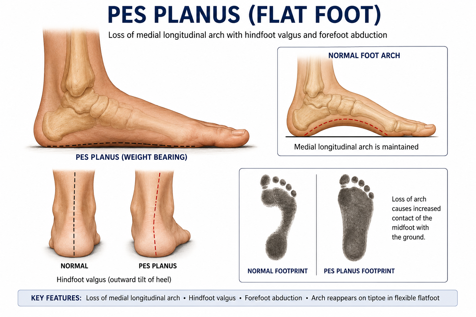
Overview — Pes Planus
Pes planus, commonly called flatfoot, is a deformity characterized by loss or reduction of the medial longitudinal arch of the foot. It is frequently associated with hindfoot valgus, forefoot abduction, and collapse of the medial column. The condition may be flexible or rigid, physiological or pathological, congenital or acquired.
Flatfoot is very common in children and is usually flexible, painless, and physiological. In adults, progressive acquired flatfoot is often related to posterior tibial tendon dysfunction, ligament attenuation, inflammatory disease, trauma, or degenerative arthritis. The key clinical task is to differentiate a benign flexible flatfoot from a painful rigid or progressive deformity requiring investigation and treatment.
- High-yield definition: Pes planus = loss of medial longitudinal arch with varying degrees of hindfoot valgus and forefoot abduction.
- Flexible flatfoot: arch is absent during standing but reappears on tiptoe or non-weight-bearing.
- Rigid flatfoot: arch remains absent even on tiptoe and is commonly associated with tarsal coalition, vertical talus, arthritis, or neuromuscular disease.
Anatomy of the Medial Longitudinal Arch
The medial longitudinal arch is the principal weight-bearing and shock-absorbing arch of the foot. It is formed by the calcaneus, talus, navicular, cuneiforms, and first three metatarsals. The talar head is the keystone of the arch and is supported inferiorly by the spring ligament and dynamically by the posterior tibial tendon.
| Structure | Role in Arch Support | Clinical Significance |
|---|---|---|
| Talus | Keystone of medial arch | Talar head plantar-medial sag causes arch collapse |
| Navicular | Insertion of posterior tibial tendon | Medial prominence and tenderness in PTT dysfunction |
| Spring ligament | Supports talar head | Attenuation contributes to adult-acquired flatfoot |
| Plantar fascia | Windlass mechanism during toe-off | Arch rises during Jack test if flexible |
| Posterior tibial tendon | Dynamic stabilizer of medial arch | Failure causes adult-acquired flatfoot |
- Exam pearl: the posterior tibial tendon is the most important dynamic stabilizer of the medial longitudinal arch.
Biomechanics of Flatfoot
Normal foot function depends on the ability of the foot to become flexible during loading and rigid during push-off. In pes planus, collapse of the medial arch disrupts this mechanism. The talus plantarflexes and adducts, the calcaneus everts into valgus, the forefoot abducts, and the medial column sags. This produces inefficient lever-arm mechanics during gait.
- Hindfoot valgus increases pronation and medial loading.
- Forefoot abduction produces the “too many toes” sign when viewed from behind.
- Medial column collapse produces midfoot sag and arch pain.
- Posterior tibial tendon failure reduces inversion power and arch support.
- Achilles tendon tightness increases midfoot collapse by forcing compensatory pronation.
- Flatfoot deformity is a three-dimensional deformity: hindfoot valgus + forefoot abduction + medial arch collapse.
Classification of Pes Planus
| Type | Key Features | Common Causes | Treatment Approach |
|---|---|---|---|
| Flexible flatfoot | Arch reappears on tiptoe or non-weight-bearing | Physiological laxity, pediatric flatfoot | Reassurance if asymptomatic; orthoses if symptomatic |
| Rigid flatfoot | Arch absent even on tiptoe; restricted subtalar motion | Tarsal coalition, vertical talus, arthritis | Investigate cause; often surgical if painful |
| Congenital flatfoot | Present early in life | Congenital vertical talus, oblique talus | Depends on rigidity and severity |
| Adult-acquired flatfoot | Progressive arch collapse in adult | Posterior tibial tendon dysfunction, ligament failure | Stage-based reconstruction or fusion |
| Neuromuscular flatfoot | Associated muscle imbalance/spasticity | Cerebral palsy, muscular dystrophy, neuropathy | Orthoses, tendon balancing, osteotomy/fusion |
Pediatric Flexible Flatfoot
Pediatric flexible flatfoot is common and usually physiological. Many children appear flat-footed because of ligamentous laxity, a thick plantar fat pad, and incomplete development of the medial arch. The arch often develops gradually with age. In an asymptomatic child with flexible painless flatfoot, treatment is usually reassurance and observation.
- Most pediatric flexible flatfeet are physiological and do not need surgery.
- The arch is absent during standing but appears on tiptoe or when the great toe is dorsiflexed.
- Pain, rigidity, asymmetry, progressive deformity, calf tightness, or recurrent ankle sprain should prompt evaluation.
- Orthoses do not permanently create an arch but may improve symptoms and shoe wear in symptomatic children.
- Achilles stretching is useful when equinus contributes to symptoms.
Rigid Flatfoot
Rigid flatfoot is more concerning than flexible flatfoot because it usually reflects structural pathology. The arch remains absent during tiptoe standing and subtalar motion is reduced. Painful rigid flatfoot in an adolescent should raise suspicion for tarsal coalition, especially calcaneonavicular or talocalcaneal coalition.
| Cause | Typical Age / Feature | Diagnostic Clue |
|---|---|---|
| Tarsal coalition | Adolescent with painful rigid flatfoot | Restricted subtalar motion, peroneal spasm, coalition on X-ray/CT |
| Congenital vertical talus | Infant with rocker-bottom foot | Fixed dorsal dislocation of navicular on talus |
| Inflammatory arthritis | Adult with painful stiff foot | Multiple joint involvement, synovitis |
| Post-traumatic arthritis | History of calcaneal, talar, or midfoot injury | Degenerative changes on weight-bearing radiographs |
Adult Acquired Flatfoot Deformity and PTT Dysfunction
Adult acquired flatfoot deformity is commonly caused by posterior tibial tendon dysfunction. The posterior tibial tendon normally inverts the hindfoot, supports the medial arch, and helps lock the transverse tarsal joints during push-off. When the tendon degenerates or ruptures, the arch collapses, the hindfoot moves into valgus, and the forefoot abducts.
- Common in middle-aged women, obesity, diabetes, hypertension, inflammatory arthritis, and steroid exposure.
- Early disease presents with pain and swelling posterior to the medial malleolus.
- Progressive disease produces medial arch collapse, too many toes sign, and inability to perform single heel rise.
- Late disease becomes rigid and arthritic.
- Adult acquired flatfoot is most commonly associated with posterior tibial tendon dysfunction.
Johnson & Strom / Myerson Classification
| Stage | Pathology | Clinical Features | Typical Management |
|---|---|---|---|
| Stage I | PTT tenosynovitis, no deformity | Medial ankle pain, tendon swelling, normal alignment | Immobilization, orthoses, physiotherapy |
| Stage II A | Flexible flatfoot, mild abduction | Arch collapse, flexible hindfoot valgus | FDL transfer + medializing calcaneal osteotomy |
| Stage II B | Flexible flatfoot with marked forefoot abduction | Too many toes sign, talonavicular uncoverage | Add lateral column lengthening if needed |
| Stage III | Rigid flatfoot with subtalar arthritis | Fixed deformity, painful stiff hindfoot | Triple arthrodesis / hindfoot fusion |
| Stage IV | Ankle valgus involvement | Deltoid insufficiency, ankle arthritis/valgus tilt | Hindfoot fusion plus ankle procedure |
Clinical Examination
Examination should be performed with the patient standing, walking, sitting, and on tiptoe. Both feet should be compared. The examiner should assess arch height, hindfoot valgus, forefoot abduction, subtalar motion, Achilles tightness, posterior tibial tendon tenderness, and neurovascular status.
| Test / Sign | How to Perform | Interpretation |
|---|---|---|
| Tiptoe test | Ask patient to stand on toes | Arch reconstitution and heel varus = flexible flatfoot |
| Single heel rise test | Patient rises on one foot | Failure suggests posterior tibial tendon dysfunction |
| Too many toes sign | Observe from behind | More lateral toes visible = forefoot abduction |
| Jack test / Hubscher maneuver | Dorsiflex great toe while standing | Arch rises if windlass mechanism intact and deformity flexible |
| Silfverskiold test | Compare ankle dorsiflexion with knee flexed and extended | Differentiates gastrocnemius tightness from tendo-Achilles tightness |
Radiographic Evaluation
Weight-bearing radiographs are essential for evaluating symptomatic flatfoot. Standard views include weight-bearing AP, lateral, and hindfoot alignment views. Radiographs assess talonavicular uncoverage, medial column sag, calcaneal pitch, talo-first metatarsal alignment, and arthritic changes.
| Measurement | Normal / Usual Value | Flatfoot Finding | Importance |
|---|---|---|---|
| Meary's angle | Around 0° | Increased plantar convexity / broken cyma line | Assesses medial arch collapse on lateral view |
| Calcaneal pitch | Approximately 17–32° | Reduced | Low pitch indicates flat arch |
| Talonavicular coverage angle | <7° commonly used | Increased | Measures forefoot abduction |
| AP talo-first metatarsal angle | Near neutral | Increased abduction angle | Assesses talar-first ray alignment |
| Hindfoot alignment view | Mild valgus/neutral | Increased valgus | Guides calcaneal osteotomy planning |
- Most asked radiological parameter in exams: Meary's angle on weight-bearing lateral foot radiograph.
Advanced Imaging
- CT is useful for suspected tarsal coalition, complex deformity, subtalar arthritis, and preoperative planning for fusion.
- MRI is useful for posterior tibial tendon degeneration, spring ligament injury, marrow edema, coalition, osteochondral pathology, and early inflammatory disease.
- Ultrasound can assess posterior tibial tendon tenosynovitis or tear dynamically but is operator-dependent.
- Rigid painful adolescent flatfoot should be investigated for tarsal coalition, commonly with CT or MRI.
Non-Operative Management
Treatment depends on symptoms, flexibility, age, etiology, and severity. Asymptomatic flexible flatfoot does not require active treatment. Symptomatic flexible flatfoot is initially treated with footwear modification, orthoses, stretching, physiotherapy, and activity modification.
- Asymptomatic pediatric flexible flatfoot = reassurance and observation.
- Footwear: supportive shoes with firm heel counter and medial arch support.
- Orthoses: medial arch support, UCBL orthosis, ankle-foot orthosis for more severe flexible deformity.
- Physiotherapy: calf stretching, posterior tibial strengthening, intrinsic foot muscle strengthening, proprioception.
- Weight reduction and activity modification help in symptomatic adult flatfoot.
- Immobilization in boot/cast may be used for painful PTT tenosynovitis.
- NSAIDs and local measures can help pain but do not correct structural deformity.
Surgical Management — Principles
Surgery is considered when pain and functional limitation persist despite adequate non-operative treatment, or when there is progressive deformity. The surgical goal is to create a plantigrade, painless, stable foot. Flexible deformities are treated with joint-sparing reconstruction, while rigid arthritic deformities generally require fusion.
| Procedure | Main Indication | Principle |
|---|---|---|
| Medializing calcaneal osteotomy | Flexible hindfoot valgus | Shifts calcaneal tuberosity medially to improve hindfoot alignment |
| FDL transfer | Posterior tibial tendon insufficiency | Replaces deficient PTT function |
| Lateral column lengthening | Forefoot abduction / talonavicular uncoverage | Lengthens lateral column to reduce abduction |
| Cotton osteotomy | Residual forefoot varus/supination after hindfoot correction | Medial cuneiform opening wedge plantarflexes medial column |
| Subtalar arthroereisis | Selected pediatric flexible flatfoot | Limits excessive subtalar pronation |
| Triple arthrodesis | Rigid flatfoot / arthritis | Fusion of subtalar, talonavicular, and calcaneocuboid joints |
Treatment of Adult Acquired Flatfoot by Stage
| Stage | Preferred Treatment | Common Surgical Options |
|---|---|---|
| Stage I | Non-operative treatment | Tenosynovectomy rarely if refractory |
| Stage II A | Joint-sparing reconstruction | Medializing calcaneal osteotomy + FDL transfer ± spring ligament repair |
| Stage II B | Reconstruction with correction of abduction | Add lateral column lengthening ± Cotton osteotomy |
| Stage III | Fusion for rigid deformity | Triple arthrodesis or double arthrodesis |
| Stage IV | Address hindfoot and ankle valgus | Triple fusion + deltoid/ankle procedure or ankle arthrodesis/arthroplasty in selected cases |
Complications
| Complication | Mechanism | Prevention / Management |
|---|---|---|
| Progressive deformity | Untreated PTT dysfunction or ligament failure | Early diagnosis, orthoses, stage-based correction |
| Lateral impingement pain | Severe hindfoot valgus causing subfibular impingement | Correct hindfoot valgus adequately |
| Overcorrection | Excessive osteotomy correction | Careful planning and intraoperative assessment |
| Nonunion | Fusion/osteotomy healing failure | Good fixation, bone graft, avoid smoking, optimize diabetes |
| Hardware irritation | Prominent screws/plates | Implant removal if symptomatic after union |
| Adjacent joint arthritis | Altered biomechanics after fusion | Joint-sparing surgery when suitable; correct alignment |
Prognosis
The prognosis depends on the type and cause of pes planus. Asymptomatic pediatric flexible flatfoot usually has an excellent natural history. Symptomatic flexible flatfoot often improves with orthoses, stretching, and activity modification. Adult acquired flatfoot is progressive if posterior tibial tendon dysfunction and ligament failure are not addressed. Early-stage disease has better outcomes with conservative care or joint-sparing reconstruction, while late rigid disease often requires arthrodesis.
- Flexible painless pediatric flatfoot: excellent prognosis.
- Painful rigid flatfoot: requires evaluation for structural pathology.
- Stage I/II adult acquired flatfoot: better outcomes with early treatment.
- Rigid arthritic flatfoot: fusion gives pain relief but sacrifices motion.
- Obesity, diabetes, smoking, inflammatory disease, and severe deformity worsen prognosis.
Exam Pearls
- Pes planus = loss of medial longitudinal arch with hindfoot valgus and forefoot abduction.
- Most pediatric flexible flatfeet are physiological and require reassurance only.
- Flexible flatfoot = arch reappears on tiptoe; rigid flatfoot = arch remains absent.
- Posterior tibial tendon is the dynamic stabilizer of the medial arch.
- Adult acquired flatfoot is commonly due to posterior tibial tendon dysfunction.
- Single heel rise test is the key clinical test for posterior tibial tendon function.
- Too many toes sign indicates forefoot abduction.
- Jack test / Hubscher maneuver demonstrates windlass mechanism and flexibility.
- Meary's angle is the most commonly asked radiographic angle.
- Rigid painful adolescent flatfoot suggests tarsal coalition.
- Triple arthrodesis = fusion of subtalar, talonavicular, and calcaneocuboid joints.
- Stage II AAFD is flexible and treated with reconstruction; Stage III is rigid and treated with fusion.
References
- Mosca VS. Flexible flatfoot in children and adolescents. J Child Orthop. 2010;4(2):107-121.
- Johnson KA, Strom DE. Tibialis posterior tendon dysfunction. Clin Orthop Relat Res. 1989;(239):196-206.
- Myerson MS. Adult acquired flatfoot deformity: treatment of dysfunction of the posterior tibial tendon. Instr Course Lect. 1997;46:393-405.
- Bluman EM, Title CI, Myerson MS. Posterior tibial tendon rupture: a refined classification system. Foot Ankle Clin. 2007;12(2):233-249.
- Deland JT. Adult-acquired flatfoot deformity. J Am Acad Orthop Surg. 2008;16(7):399-406.
- Vulcano E, Deland JT, Ellis SJ. Approach and treatment of the adult acquired flatfoot deformity. Curr Rev Musculoskelet Med. 2013;6(4):294-303.
- Staheli LT. Planovalgus foot deformity. Current status. J Am Podiatr Med Assoc. 1999;89(2):94-99.