
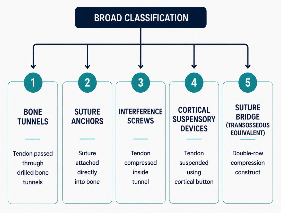
Overview — Tendon-to-Bone Fixation
Tendon-to-bone fixation is a core principle in orthopaedic surgery, sports medicine, and reconstructive procedures. It refers to the mechanical attachment of a tendon, ligament graft, or soft tissue structure to bone until biological healing occurs at the tendon-bone interface. It is used in rotator cuff repair, biceps tenodesis, ACL and PCL reconstruction, collateral ligament reconstruction, patellar tendon repair, quadriceps tendon repair, distal biceps repair, Achilles insertion repair, ankle ligament reconstruction, and multiple tendon transfer procedures.
The ideal fixation method should provide sufficient initial strength, resist cyclic loading, maintain tendon-to-bone contact, minimize gap formation, allow biological incorporation, and permit safe rehabilitation. The choice of fixation depends on anatomical site, tendon quality, bone quality, direction of pull, expected loads, available bone stock, and whether the construct is intra-articular or extra-articular.
- High-yield definition: tendon-to-bone fixation = mechanical attachment of tendon/graft to bone using sutures, anchors, tunnels, screws, buttons, staples, washers, or suspensory devices until biological healing occurs.
- Mechanical fixation is immediate; biological healing is gradual and takes weeks to months.
- Failure may occur by suture pullout, anchor pullout, screw divergence, graft slippage, bone tunnel widening, or biological non-healing.
Biology of Tendon-to-Bone Healing
Native tendon insertion is a specialized enthesis with a gradual transition from tendon to unmineralized fibrocartilage, mineralized fibrocartilage, and bone. This transition decreases stress concentration. Surgical repair rarely recreates this complex native enthesis perfectly; healing usually occurs through fibrovascular scar tissue that gradually matures and mineralizes.
| Phase | Time Period | Biological Events | Clinical Relevance |
|---|---|---|---|
| Inflammatory phase | First few days | Hematoma, inflammatory cell recruitment, cytokine release | Fixation construct carries most of the load |
| Proliferative phase | 1–6 weeks | Fibroblast proliferation, collagen deposition, vascular ingrowth | Controlled loading may help; excessive load causes gap formation |
| Remodelling phase | 6 weeks to months | Collagen organization, mineralization, interface maturation | Gradual strengthening and functional rehabilitation |
- Exam pearl: tendon-to-bone healing is initially weak and depends on fixation strength; biological incorporation becomes more important after the early postoperative period.
Mechanical Principles of Fixation
A tendon-to-bone construct must resist tensile load, shear, cyclic displacement, and gap formation. The fixation should compress the tendon against a bleeding bone bed and maintain contact during early motion. Mechanical strength depends on suture material, stitch configuration, anchor design, bone quality, tunnel diameter, screw size, graft diameter, and direction of loading.
- Initial fixation strength must exceed early rehabilitation forces.
- Contact area and compression improve biological healing potential.
- Gap formation weakens healing and increases risk of failure.
- Bone quality is critical for anchors, screws, staples, and cortical buttons.
- Tendon quality determines whether sutures cut through the tissue.
- The weakest link may be tendon-suture interface, anchor-bone interface, graft-screw interface, cortical button-bone interface, or biological tendon-bone interface depending on the procedure.
Common Tendon-to-Bone Fixation Methods
| Method | Principle | Common Examples | Main Limitation |
|---|---|---|---|
| Bone tunnels | Sutures or graft passed through transosseous tunnels and tied over bone bridge | Patellar tendon repair, quadriceps tendon repair, transosseous rotator cuff repair | Bone bridge fracture, technical demand |
| Suture anchors | Anchor fixed into bone with sutures attached to tendon | Rotator cuff repair, Bankart repair, Achilles insertion repair | Anchor pullout in weak bone |
| Interference screws | Screw compresses graft against bone tunnel wall | ACL/PCL reconstruction, biceps tenodesis | Graft laceration, screw divergence, tunnel widening |
| Cortical buttons | Button flips on far cortex and suspends graft/tendon | ACL femoral fixation, distal biceps repair | Loop elongation, cortical breach, button malposition |
| Staples / washers | External compression of tendon/graft to bone | MCL reconstruction, extra-articular tenodesis, tibial graft fixation | Prominence and irritation |
Bone Tunnel Fixation
Bone tunnel fixation is one of the oldest and most reliable tendon-to-bone fixation methods. Drill holes are made through bone, sutures are passed through the tendon and tunnels, and the tendon is pulled down to the prepared bone surface. The sutures are tied over a bony bridge or through the opposite cortex. This produces broad tendon-bone contact and does not require expensive implants.
- Principle: transosseous sutures convert tensile force in the tendon into compression of tendon against a bleeding bone bed.
- Common in patellar tendon repair and quadriceps tendon repair through patellar drill holes.
- Rotator cuff transosseous repair uses tunnels through the greater tuberosity.
- Advantages: low cost, broad contact area, no implant-related imaging artefact.
- Disadvantages: technically demanding, risk of tunnel convergence, bony bridge fracture, and difficulty in osteoporotic bone.
Suture Anchor Fixation
Suture anchors are implants inserted into bone with attached sutures that are passed through tendon or ligament and tied or locked to secure soft tissue against bone. They may be metallic, bioabsorbable, PEEK, biocomposite, or all-suture anchors. They are widely used in arthroscopic and open soft tissue repairs because they simplify fixation and avoid long transosseous tunnels.
| Anchor Type | Features | Advantages | Limitations |
|---|---|---|---|
| Metal anchor | Titanium or stainless steel | High strength, visible on X-ray | MRI artefact, difficult revision, cartilage damage if prominent |
| Bioabsorbable anchor | PLLA/PLGA or biocomposite | Less permanent hardware | Cyst, inflammatory reaction, breakage |
| PEEK anchor | Inert polymer | Radiolucent, MRI compatible, no resorption reaction | Permanent implant |
| All-suture anchor | Soft anchor that expands under cortex | Small drill hole, preserves bone stock | Technique sensitive, depends on cortical bone quality |
| Knotless anchor | Locks suture without arthroscopic knot | Low profile, faster, useful in double-row repairs | Tension must be set correctly before final locking |
Interference Screw Fixation
Interference screw fixation is most commonly used for ligament reconstruction and tendon graft fixation inside bone tunnels. The screw is inserted between the graft and tunnel wall. As the screw advances, it compresses the graft against cancellous or cortical bone, creating frictional fixation and promoting tendon-to-bone healing within the tunnel.
- Principle: interference screw fixation works by compressing the graft against the bone tunnel wall, producing friction and contact pressure.
- Used in ACL reconstruction with hamstring graft, BPTB graft, quadriceps tendon graft, and PCL reconstruction.
- Also used in biceps tenodesis and some tendon transfer procedures.
- Screw diameter is commonly matched to tunnel size or slightly larger depending on graft type and bone quality.
- Metal screws provide strength but create MRI artefact and revision difficulty.
- Bioabsorbable and biocomposite screws reduce permanent hardware but may break or cause inflammatory cysts.
Cortical Button and Suspensory Fixation
Cortical button fixation uses a small metallic button passed through a bone tunnel and flipped on the far cortex. The tendon or graft is suspended from the button using sutures or adjustable loop systems. This method provides strong fixation by relying on cortical bone rather than cancellous bone.
| Application | Example | Reason for Use |
|---|---|---|
| ACL femoral fixation | Endobutton / adjustable loop device | Strong cortical fixation, useful with hamstring grafts |
| Distal biceps repair | Button through radial tuberosity | High load-to-failure and secure tendon docking |
| PCL reconstruction | Suspensory cortical fixation | Useful with graft loops and tunnels |
- Advantages: strong fixation, small implant, useful in cortical bone, commonly used arthroscopically.
- Limitations: button malposition, soft tissue interposition, tunnel widening, loop elongation, and graft motion within tunnel.
- Cortical buttons are suspensory fixation devices; interference screws are aperture compression devices.
Suture Configuration and Tendon-Suture Interface
Even when bone fixation is strong, failure may occur at the tendon-suture interface. Tendon quality, number of suture limbs, bite depth, locking configuration, and suture material influence strength. Locking stitches distribute load more effectively and resist pullout better than simple stitches.
| Suture Pattern | Principle | Common Use | Key Point |
|---|---|---|---|
| Simple stitch | Single pass through tendon | Small soft tissue repairs | Lower pullout strength |
| Mattress stitch | Broader compression across tissue | Rotator cuff, tendon repair | Distributes load better than simple stitch |
| Krackow stitch | Locking running stitch along tendon | Patellar tendon, quadriceps tendon, Achilles repair | High tendon purchase and pullout resistance |
| Mason-Allen stitch | Horizontal mattress with locking loop | Rotator cuff repair | Strong tissue grasp in cuff tendon |
| Rip-stop configuration | Reinforcing suture prevents tissue cut-through | Poor-quality cuff or degenerative tendon | Useful when tendon quality is weak |
Rotator Cuff Repair
Rotator cuff repair is one of the most common clinical examples of tendon-to-bone fixation. The torn cuff tendon is mobilized and repaired back to the greater tuberosity footprint using suture anchors, transosseous tunnels, or transosseous-equivalent double-row constructs. Biological healing depends on tendon quality, footprint preparation, compression, and controlled rehabilitation.
- Single-row repair: anchors placed near articular margin; simpler and less expensive.
- Double-row repair: medial and lateral row anchors improve footprint contact area and compression.
- Transosseous-equivalent / suture bridge: medial row sutures crossed and fixed laterally to compress tendon broadly.
- Transosseous repair: sutures passed through bone tunnels without anchors.
- Key concept: rotator cuff healing depends on stable tendon compression over the greater tuberosity footprint.
ACL Reconstruction Fixation
In ACL reconstruction, tendon-to-bone healing occurs within femoral and tibial bone tunnels. Fixation methods vary according to graft type. Bone-patellar tendon-bone graft heals partly by bone-to-bone healing at the bone plug interface, whereas hamstring and quadriceps soft tissue grafts require tendon-to-bone incorporation within the tunnel.
| Graft Type | Common Fixation | Healing Characteristic |
|---|---|---|
| BPTB graft | Interference screws for bone plugs | Bone-to-bone healing, relatively faster incorporation |
| Hamstring graft | Femoral cortical button, tibial interference screw/post | Tendon-to-bone healing within tunnel |
| Quadriceps tendon graft | Button, screw, or hybrid fixation | Soft tissue or bone plug dependent |
- Femoral fixation often uses cortical button or interference screw.
- Tibial fixation is commonly the weaker side due to lower bone density and tunnel direction.
- Hybrid fixation combines aperture fixation with backup cortical/post fixation.
Biceps, Extensor Mechanism, and Foot-Ankle Examples
| Procedure | Fixation Methods | Important Point |
|---|---|---|
| Proximal biceps tenodesis | Interference screw, suture anchor, cortical button, soft tissue tenodesis | Avoid overtensioning and persistent groove pain |
| Distal biceps repair | Cortical button, interference screw, suture anchors, bone tunnels | Cortical button fixation has high load-to-failure |
| Patellar tendon repair | Krackow sutures through patellar bone tunnels or anchors | Prevent gap formation during knee flexion |
| Quadriceps tendon repair | Patellar tunnels or suture anchors at superior pole | Strong fixation needed for early controlled motion |
| Achilles insertion repair | Calcaneal anchors, double-row bridge | Footwear irritation and wound healing matter |
| Brostrom repair | Suture anchors in distal fibula | Restores ATFL/CFL attachment |
Factors Affecting Fixation Choice
| Factor | Effect on Fixation | Example |
|---|---|---|
| Bone quality | Poor bone increases anchor/screw pullout | Osteoporotic greater tuberosity may need larger/multiple anchors |
| Tendon quality | Degenerative tendon may fail by suture cut-through | Massive cuff tear may need rip-stop or margin convergence |
| Direction of pull | Fixation strongest when load is aligned with construct design | Interference screw divergence weakens ACL fixation |
| Footprint size | Larger footprint may require double-row or multiple fixation points | Rotator cuff repair |
| Revision potential | Bone preservation and imaging matter | All-suture/PEEK anchors may be preferred in selected cases |
Complications and Failure Mechanisms
| Failure / Complication | Mechanism | Common Scenario | Prevention |
|---|---|---|---|
| Anchor pullout | Poor bone purchase or wrong insertion angle | Osteoporotic cuff repair | Correct anchor size, good bone bed, avoid over-tensioning |
| Suture cut-through | Suture slices through weak tendon | Degenerative cuff or chronic tendon rupture | Locking/rip-stop sutures, wider bites, augmentation |
| Graft slippage | Inadequate tunnel compression or fixation | ACL hamstring graft tibial side | Correct screw size, backup fixation, graft tensioning |
| Tunnel widening | Micromotion, synovial fluid ingress, biological reaction | ACL reconstruction | Anatomic tunnel, aperture fixation, minimize graft motion |
| Hardware irritation | Prominent staple, washer, screw, or button | Extra-articular ligament fixation | Low-profile implants, proper placement |
| Biological failure | Poor tendon-bone healing despite intact fixation | Massive cuff tear, smoker, diabetes | Optimize biology, footprint preparation, protected rehab |
Rehabilitation Principles
Rehabilitation after tendon-to-bone fixation must balance protection of the healing interface with prevention of stiffness, adhesions, and muscle atrophy. Biological healing is slow, so early aggressive loading can cause gap formation or failure even when intraoperative fixation appears strong.
- Early phase: protect repair, control pain/swelling, maintain adjacent joint motion.
- Intermediate phase: begin controlled passive or active-assisted motion depending on procedure.
- Strengthening phase: progressive loading only after adequate biological healing.
- Return-to-sport phase: sport-specific strengthening, proprioception, functional testing.
- Rehabilitation depends on biology, not just implant strength: a strong construct still needs time for tendon-to-bone healing.
Comparison of Fixation Methods
| Method | Strength | Cost | Best Use | Main Concern |
|---|---|---|---|---|
| Bone tunnels | Good if bone bridge strong | Low | Extensor mechanism, transosseous cuff | Technical demand |
| Suture anchors | Good to excellent | Moderate/high | Arthroscopic repairs | Anchor pullout |
| Interference screws | Excellent aperture fixation | Moderate | ACL/PCL tunnels, tenodesis | Graft damage or slippage |
| Cortical buttons | Very high cortical fixation | Moderate/high | ACL femur, distal biceps | Suspensory micromotion |
| Staples/washers | Good backup fixation | Low/moderate | Extra-articular grafts | Prominence and irritation |
Exam Pearls
- Tendon-to-bone healing occurs through fibrovascular scar rather than perfect recreation of the native enthesis.
- Native enthesis has four zones: tendon, unmineralized fibrocartilage, mineralized fibrocartilage, and bone.
- Mechanical fixation is immediate; biological fixation develops over weeks to months.
- Bone tunnels are low-cost and useful in patellar tendon, quadriceps tendon, and transosseous cuff repairs.
- Suture anchors are common in rotator cuff, Bankart, SLAP, Achilles insertion, and Brostrom repairs.
- Interference screw = aperture fixation by compressing graft against tunnel wall.
- Cortical button = suspensory fixation relying on far cortex.
- BPTB ACL graft has bone-to-bone healing; hamstring ACL graft has tendon-to-bone healing.
- Tibial side fixation in ACL reconstruction is often weaker than femoral side fixation.
- Rotator cuff repair aims to restore tendon contact over greater tuberosity footprint.
- Failure may occur at tendon-suture interface, implant-bone interface, graft-tunnel interface, or biological interface.
- Rehabilitation should respect biological healing time, not only implant strength.
References
- Rodeo SA, Arnoczky SP, Torzilli PA, Hidaka C, Warren RF. Tendon-healing in a bone tunnel: a biomechanical and histological study in the dog. J Bone Joint Surg Am. 1993;75(12):1795-1803.
- Thomopoulos S, Williams GR, Soslowsky LJ. Tendon to bone healing: biomechanical, structural, and compositional properties. J Biomech Eng. 2003;125(1):106-113.
- Benjamin M, Toumi H, Ralphs JR, Bydder G, Best TM, Milz S. Where tendons and ligaments meet bone: attachment sites or entheses. J Anat. 2006;208(4):471-490.
- Burkhart SS, Lo IKY. Arthroscopic rotator cuff repair. J Am Acad Orthop Surg. 2006;14(6):333-346.
- Mazzocca AD, Bicos J, Arciero RA, Romeo AA, Cohen MS, Nicholson GP. Biomechanical evaluation of distal biceps tendon repair techniques. Am J Sports Med. 2007;35(2):252-258.
- Milano G, Mulas PD, Ziranu F, Piras S, Manunta A, Fabbriciani C. Comparison between femoral fixation devices for ACL reconstruction. Arthroscopy. 2006;22(6):660-668.