
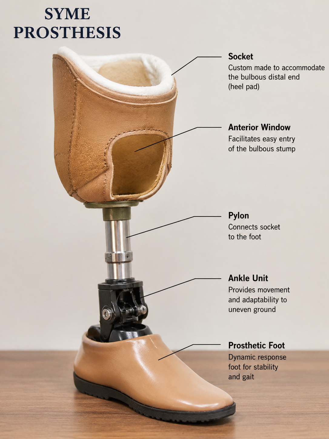
Overview — Syme's Amputation and Syme's Prosthesis
Syme's amputation is an ankle disarticulation procedure in which the foot is removed through the ankle joint while preserving the heel pad as a durable end-bearing surface. It occupies an important position between partial foot amputations and transtibial amputations because it preserves limb length, provides a broad weight-bearing stump, and allows more efficient gait than more proximal amputations when performed and fitted correctly.
The classical operation was described by James Syme in 1843. In modern orthopaedic practice, Syme's amputation is considered for selected patients with non-salvageable foot pathology, especially when the heel pad is viable and the posterior tibial artery supply is intact. The major technical challenge is maintaining the heel pad directly beneath the distal tibia. The major prosthetic challenge is accommodating the bulbous distal end while still providing a cosmetically acceptable and mechanically efficient prosthesis.
- High-yield definition: Syme's amputation = ankle disarticulation with removal of the foot, excision of malleoli, and preservation/fixation of the heel pad to create an end-bearing stump.
- It is a true end-bearing amputation level, unlike the standard transtibial amputation where most load is transmitted through the socket walls.
- Success depends on vascularity, infection control, heel pad stability, and appropriate prosthetic design.
Historical Background
James Syme introduced the ankle disarticulation technique as a limb-preserving alternative to more proximal amputations. The concept was important because it preserved the patient's leg length and used the naturally weight-bearing heel pad as the terminal surface. Later surgeons modified the operation to improve heel pad stability, prosthetic fitting, and function. Although its popularity decreased with advances in transtibial prosthetics, Syme's amputation remains a valuable option in carefully selected patients.
- Original principle: remove the foot through the ankle joint and bring the heel pad under the distal tibia.
- Modern principle: create a stable, painless, plantigrade, end-bearing stump that can tolerate prosthetic loading.
- Commonly compared in examinations with Boyd, Pirogoff, Chopart, and below-knee amputation.
Surgical Anatomy
The surgical success of Syme's amputation depends on understanding the heel flap, posterior tibial vascularity, ankle joint anatomy, malleoli, and terminal branches of the major nerves. The heel pad is a specialized weight-bearing structure composed of thick glabrous skin, fibrous septae, and fat compartments. It is designed to tolerate compressive load during stance phase.
| Structure | Importance | Surgical Relevance |
|---|---|---|
| Heel pad | Specialized end-bearing tissue | Must be preserved and fixed under distal tibia |
| Posterior tibial artery | Major blood supply to heel flap | Integrity is critical for flap healing |
| Malleoli | Medial and lateral ankle prominences | Removed or bevelled to reduce bulbosity and improve prosthetic fit |
| Tibial nerve | Posterior neurovascular bundle | Divided proximally under traction to reduce neuroma pain |
| Achilles tendon | Posterior tendon insertion into calcaneus | Detached with posterior flap during foot removal |
- Exam pearl: posterior tibial artery supply to the heel flap is the key vascular requirement for Syme's amputation.
Biomechanical Principles
Syme's amputation preserves nearly the full length of the tibial lever arm. The patient can bear weight through the distal stump because the heel pad is placed beneath the tibia. This makes gait more energy efficient than a transtibial amputation and allows limited standing or transfers without a prosthesis in selected patients. However, the bulbous distal stump makes prosthetic fitting more difficult than transtibial amputation.
- The main biomechanical advantage of Syme's amputation is true end-bearing through the preserved heel pad.
- Long lever arm improves control of the prosthesis and reduces energy expenditure.
- End-bearing decreases dependence on proximal socket suspension and reduces pressure over the patellar tendon region.
- The bulbous distal end improves suspension but creates cosmetic and donning challenges.
- Medial-lateral instability or heel pad migration compromises prosthetic comfort and gait.
Indications
| Indication | Examples | Why Syme May Be Useful |
|---|---|---|
| Trauma | Crush foot, mangled forefoot/midfoot, non-reconstructable foot injury | Preserves limb length and end-bearing surface if heel is intact |
| Infection | Forefoot/midfoot osteomyelitis, severe soft tissue infection sparing heel | Allows removal of infected foot while preserving durable stump |
| Diabetic foot | Selected cases with adequate perfusion and viable heel pad | Can provide end-bearing stump but requires careful vascular selection |
| Congenital deformity | Severe foot deficiency or deformity not suitable for reconstruction | Maintains limb length in children and allows durable prosthetic fitting |
| Tumour | Selected distal foot lesions where oncologic margins permit | Limb-sparing level if heel and ankle region are not involved |
Contraindications
| Contraindication | Reason | Practical Implication |
|---|---|---|
| Non-viable heel pad | Loss of end-bearing tissue | Syme stump will fail or ulcerate |
| Posterior tibial artery compromise | Heel flap vascularity at risk | High risk of flap necrosis |
| Severe peripheral vascular disease | Poor healing potential | Consider more proximal level if perfusion inadequate |
| Infection involving heel | Cannot preserve safe heel pad | Contraindicates heel-pad-based end-bearing stump |
| Severe neuropathic heel | Insensate end-bearing surface | Ulceration and breakdown risk |
Advantages and Disadvantages
| Advantages | Disadvantages |
|---|---|
| End-bearing stump with preserved heel pad | Bulbous distal stump makes prosthetic fitting difficult |
| Long lever arm and better proprioceptive control | Inferior cosmesis compared with transtibial prosthesis |
| Lower energy expenditure than transtibial amputation | Risk of heel pad migration |
| Patient may stand/transfer short distances without prosthesis | Requires viable posterior heel flap and good vascularity |
| Useful in selected children and active patients | Can be difficult in diabetic/dysvascular patients if selection is poor |
Preoperative Evaluation
Preoperative assessment determines whether a Syme level is likely to heal and function. The surgeon must assess infection extent, heel pad quality, vascular supply, soft tissue viability, neuropathy, patient function, and prosthetic feasibility. In traumatic cases, the heel must be intact or reconstructable. In diabetic and dysvascular cases, vascular assessment is critical.
- Assess posterior heel skin: viability, sensation, ulceration, infection, scarring, and soft tissue thickness.
- Assess vascularity: posterior tibial pulse, Doppler signals, ABI/toe pressures, TcPO2 or vascular imaging when required.
- Evaluate infection: clinical extent, radiographs, MRI if osteomyelitis extent is uncertain, and culture-guided antibiotic planning.
- Assess patient factors: diabetes control, nutrition, renal disease, smoking, mobility goals, ability to use prosthesis, and rehabilitation support.
- Do not choose Syme's amputation merely to preserve length if the heel pad is non-viable or vascularity is inadequate.
Classical Surgical Technique
The operation aims to disarticulate the ankle, remove the foot, excise the malleoli, and secure the heel pad centrally beneath the distal tibia. The posterior heel flap is fashioned carefully so that the heel pad remains vascular, stable, and positioned for end-bearing.
| Step | Technique | Key Point |
|---|---|---|
| 1. Flap planning | Create anterior incision across ankle and posterior heel flap | Posterior flap must include durable heel pad |
| 2. Ankle disarticulation | Open ankle joint and remove foot through tibiotalar joint | Avoid unnecessary trauma to heel flap |
| 3. Malleolar removal | Excise medial and lateral malleoli flush with distal tibia/fibula | Reduces bulbosity and improves prosthetic fit |
| 4. Nerve management | Traction neurectomy of tibial, superficial/deep peroneal, sural, and saphenous nerves | Allow nerves to retract proximally away from pressure areas |
| 5. Heel pad fixation | Secure heel pad to distal tibia/anterior soft tissue/periosteum | Most important step to prevent migration |
| 6. Closure | Tension-free closure with drain if needed and compressive dressing | Avoid dog ears, pressure points, and flap tension |
- Most important technical principle: stable central fixation of the heel pad beneath the distal tibia.
Heel Pad Preservation and Fixation
Heel pad migration is the classic complication of Syme's amputation. If the heel pad migrates posteriorly, laterally, or medially, the patient loses the stable end-bearing surface and develops pain, ulceration, prosthetic difficulty, or need for revision. Therefore, the heel pad must be mobilized carefully and fixed securely.
- Preserve the posterior heel flap with adequate soft tissue and vascularity.
- Avoid excessive thinning or devascularization of the heel pad.
- Position the heel pad directly under the distal tibial weight-bearing surface.
- Fixation methods include suturing the heel pad/fascia to anterior tibial periosteum, drill holes in distal tibia, tendon/periosteal anchorage, or myodesis-like soft tissue fixation.
- Bevel distal tibial and fibular prominences to improve soft tissue seating.
- Heel pad stability is the most important determinant of a comfortable end-bearing Syme stump.
Modifications of Syme's Amputation
Several modifications of Syme's amputation were developed to improve heel pad stability, preserve length, improve prosthetic fitting, or provide a broader bony end-bearing surface. Some are true modifications of the Syme concept, while others are related hindfoot amputations that are commonly discussed alongside Syme's amputation in orthopaedic examinations.
| Modification / Related Procedure | Principle | Advantage | Limitation |
|---|---|---|---|
| Classical Syme with malleolar excision | Ankle disarticulation with removal of malleoli and heel pad fixation | End-bearing stump with reduced distal prominence | Heel pad migration if fixation fails |
| Two-stage Syme | Initial guillotine/disarticulation for infection, later definitive revision and closure | Useful when infection control is needed before final stump formation | Requires second procedure and prolonged treatment |
| Sarmiento-type modification | Emphasizes contouring of distal tibia/fibula and secure heel pad stabilization | Improves prosthetic fit and reduces migration | Requires meticulous soft tissue handling |
| Boyd amputation | Calcaneus is retained and fused to distal tibia after talus removal | Less heel pad migration; broad bony end-bearing surface | Longer bulbous stump; risk of nonunion; difficult prosthetic cosmesis |
| Pirogoff amputation | Posterior calcaneus is rotated and fused to distal tibia | Preserves heel pad attached to calcaneal bone; strong end-bearing | Technically demanding; fusion required; bulky stump |
| Modified Pirogoff / calcaneotibial fusion variants | Different osteotomy orientations and fixation methods to improve alignment and fusion | Can improve durable end-bearing in selected cases | Requires viable calcaneus and reliable union |
- Exam comparison: Syme preserves the heel pad alone; Boyd preserves the calcaneus and fuses it to tibia; Pirogoff rotates part of calcaneus and fuses it to tibia.
- Boyd and Pirogoff procedures reduce heel pad migration because the heel pad remains attached to calcaneal bone, but they create a bulkier stump and require bony union.
- Two-stage Syme is especially relevant in infected diabetic foot cases where definitive closure is unsafe at the first operation.
Comparison with Other Amputation Levels
| Feature | Syme | Transtibial / BKA | Partial Foot |
|---|---|---|---|
| End-bearing | Yes | Partial / socket-dependent | Variable |
| Limb length | Excellent preservation | Shorter | Best preservation |
| Energy expenditure | Lower than BKA | Higher than Syme | May be efficient if stable |
| Prosthetic cosmesis | Less cosmetic due to bulbous stump | Better cosmetic options | Shoe filler/AFO may be cosmetic |
| Risk of ulceration | Heel pad migration/socket pressure | Socket pressure areas | High if equinus/imbalance develops |
| Walking without prosthesis | Possible for short transfers in selected patients | Difficult | Often possible depending on level |
Postoperative Care
Postoperative care aims to protect the heel flap, prevent migration, control swelling, maintain knee and hip mobility, and prepare the stump for prosthetic fitting. Weight-bearing is delayed until the wound has healed and the heel pad is stable.
- Apply a compressive dressing or rigid dressing to control edema and maintain heel pad position.
- Monitor flap vascularity closely, especially in diabetic and dysvascular patients.
- Keep limb elevated and avoid pressure over the posterior flap.
- Begin hip and knee range-of-motion exercises early.
- Delay prosthetic fitting until soft tissue swelling reduces and wound healing is complete.
- Use temporary/provisional prosthesis before definitive socket once stump volume stabilizes.
Complications
| Complication | Mechanism | Prevention / Management |
|---|---|---|
| Heel pad migration | Inadequate fixation or soft tissue instability | Secure fixation; revision fixation or higher amputation if severe |
| Flap necrosis | Poor vascularity, posterior tibial artery compromise, excessive tension | Careful vascular selection, tension-free closure |
| Infection | Residual infection, diabetic wound risk, hematoma | Debridement, antibiotics, staged surgery if needed |
| Ulceration | Socket pressure, migrated heel pad, insensate stump | Socket adjustment, offloading, skin care, revision if recurrent |
| Neuroma pain | Terminal nerve ends in pressure area | Traction neurectomy; revision neuroma surgery if persistent |
| Poor prosthetic fit | Bulbous stump, edema, bony prominence, unstable heel pad | Experienced prosthetist, window socket, socket modification |
Syme's Prosthesis — Principles
A Syme prosthesis must allow entry of the bulbous distal stump, transmit load through the end-bearing heel pad, provide mediolateral stability, and restore foot length for gait. Because the distal end is wider than the narrow supramalleolar region, donning the prosthesis requires special socket design.
- Core prosthetic problem: the bulbous distal stump improves suspension but makes socket entry difficult.
- Socket must allow distal end-bearing while avoiding pressure over bony prominences and scar.
- A removable medial window, posterior opening, split socket, or flexible inner liner may be used to allow entry.
- Prosthetic foot selection depends on patient activity level, limb length clearance, cosmesis, and available build height.
- Modern designs may use carbon composite, silicone liners, energy-storing feet, and low-profile components.
Components of Syme's Prosthesis
| Component | Function | Special Point in Syme Prosthesis |
|---|---|---|
| Socket | Encloses stump and transmits load | Must accommodate bulbous distal end |
| Window / door | Allows stump entry into socket | Commonly medial removable window |
| Distal end-bearing area | Supports body weight through heel pad | Must be well-contoured and cushioned |
| Pylon / shank | Connects socket to foot | Limited space due to long residual limb |
| Prosthetic foot | Provides stance stability and rollover | Requires low-profile design because of reduced build height |
| Suspension | Retains prosthesis during swing phase | Bulbous stump can provide self-suspension with appropriate socket |
Socket Designs
Traditional Syme prostheses used a hard socket with a removable medial window to allow the bulbous stump to pass into the narrower part of the socket. Modern designs may use flexible inner sockets, laminated outer frames, silicone liners, or posterior opening designs. The choice depends on stump shape, skin condition, activity level, and available prosthetic expertise.
| Socket Type | Advantage | Limitation |
|---|---|---|
| Medial window socket | Allows entry of bulbous stump; classic design | Window may be bulky or cosmetically visible |
| Posterior opening socket | Improves cosmesis in some designs | Must avoid posterior flap pressure |
| Flexible inner socket | Improves comfort and donning | Requires skilled fabrication |
| Silicone liner system | Skin protection and suspension | Heat, sweating, and liner maintenance issues |
Rehabilitation
Rehabilitation begins before surgery with counselling and continues through wound healing, stump shaping, prosthetic training, and gait retraining. The patient must learn skin inspection, stump hygiene, progressive loading, and safe ambulation.
- Preoperative counselling: realistic expectations, prosthetic options, potential need for revision, and long-term skin care.
- Early postoperative phase: edema control, pain control, wound monitoring, knee/hip exercises, and transfer training.
- Pre-prosthetic phase: stump shaping, desensitization, scar care, strengthening, and balance training.
- Prosthetic phase: gradual end-bearing, gait training, stair training, endurance training, and socket adjustments.
- Long-term care: daily skin inspection, especially in diabetics and neuropathic patients.
Pediatric and Diabetic Foot Considerations
In children, Syme's amputation may be useful for congenital deficiency or severe deformity because it preserves limb length and provides a durable end-bearing stump. However, growth, prosthetic replacement, and cosmesis must be considered. In diabetic patients, careful selection is essential because wound failure, infection, vascular disease, and neuropathy can compromise outcomes.
- Pediatric Syme: good end-bearing, preserved knee function, and easier prosthetic control; socket changes are needed with growth.
- Diabetic Syme: consider only if heel pad is viable, perfusion is adequate, infection is controlled, and patient can perform skin care.
- Neuropathy increases risk of unrecognized ulceration.
- Two-stage Syme may be safer when infection control is uncertain.
- In diabetic foot, vascularity and heel pad quality are more important than simply preserving amputation length.
Functional Outcomes
Functional outcome after Syme's amputation can be excellent in selected patients. Energy cost is generally lower than transtibial amputation because the knee is preserved, the limb lever arm is long, and load can be transmitted through the distal stump. Many patients achieve community ambulation with a well-fitted prosthesis. Poor outcomes are usually related to heel pad migration, wound complications, vascular insufficiency, prosthetic difficulty, or severe comorbidity.
- Best candidates: intact heel pad, good vascularity, preserved knee function, motivated patient, and access to skilled prosthetics.
- Good outcomes: painless end-bearing, stable heel pad, well-fitting prosthesis, independent gait.
- Poor prognostic factors: vascular disease, severe neuropathy, active infection, unstable heel pad, poor compliance, and repeated ulceration.
Exam Pearls
- Syme's amputation = ankle disarticulation with preservation of heel pad for end-bearing.
- Described by James Syme in 1843.
- Most important structure to preserve = heel pad with posterior tibial artery supply.
- Most important complication = heel pad migration.
- Malleoli are removed or bevelled to reduce distal bulbosity and improve prosthetic fit.
- Major advantage over BKA = true end-bearing with lower energy expenditure and longer lever arm.
- Major disadvantage = bulbous stump and difficult prosthetic cosmesis.
- Syme prosthesis often requires a removable medial window or flexible socket for stump entry.
- Boyd amputation = talus removed, calcaneus fused to tibia.
- Pirogoff amputation = posterior calcaneus rotated and fused to tibia.
- Two-stage Syme may be useful in infection.
- In diabetic foot, adequate vascularity and viable heel pad are mandatory.
- Patient may stand briefly without a prosthesis because the stump is end-bearing.
References
- Syme J. Amputation at the ankle joint. Original description, 1843.
- Saini UC, Saini ML. Syme's Amputation: Do We Need It in 2020? Journal of Foot and Ankle Surgery Asia Pacific. 2021.
- Orthobullets. Amputations: Syme amputation technique and principles. Updated 2026.
- Wheeless' Textbook of Orthopaedics. Syme's Amputation: indications, technique, and complications.
- O&P Virtual Library. The Syme Ankle Disarticulation: Prosthetic Management.
- O&P Virtual Library. Syme's Prosthesis: Prosthetic fitting and fabrication principles.
- Andronic O, et al. Modifications of the Pirogoff amputation technique in adults. Journal of Clinical Orthopaedics and Trauma. 2020.
- Campbell JT. Syme's, Boyd's, Chopart's, and Pirogoff's amputations. Foot and Ankle Clinics. 1999.
- Image - AI-Generated